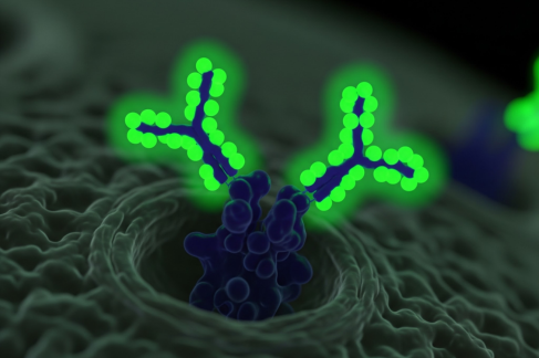
Fluorescent secondary antibodies have become indispensable tools in cellular and molecular biology, particularly in the study of cellular signaling pathways. By enabling precise localization, quantification, and interaction analysis of proteins, these antibodies provide insights into the dynamic processes that govern cellular function and disease progression.
Understanding Secondary Antibodies
Secondary antibodies are designed to bind specifically to primary antibodies, which are directed against a target antigen. Conjugated to fluorescent dyes, these secondary antibodies emit light upon excitation, allowing visualization under a fluorescence microscope. This indirect detection method amplifies the signal, enhancing sensitivity and enabling the study of low-abundance proteins.
Fluorescence Microscopy: A Window into Cellular Signaling
Fluorescence microscopy is a powerful technique that utilizes the emission of light from fluorophores to visualize cellular structures and processes. When secondary antibodies are conjugated to fluorophores, they bind to primary antibodies attached to target proteins. The emitted fluorescence allows researchers to observe the spatial and temporal dynamics of signaling molecules within cells.
Amplification of Signal for Enhanced Sensitivity
One of the key advantages of using a fluorescence secondary antibody is signal amplification. Multiple secondary antibodies can bind to a single primary antibody, increasing the overall fluorescence intensity. This amplification is particularly beneficial when detecting low-abundance proteins or subtle changes in signaling pathways.
Studying Protein Localization and Expression
Secondary antibody fluorescence enables the precise localization of proteins within cellular compartments. By tagging primary antibodies with fluorescently labeled secondary antibodies, researchers can map the distribution of signaling proteins, such as kinases and phosphatases, to specific cellular regions. This spatial information is crucial for understanding how signaling pathways are compartmentalized and regulated.
Investigating Protein-Protein Interactions
Fluorescent secondary antibodies facilitate the study of protein-protein interactions, a fundamental aspect of cellular signaling. Techniques like Förster resonance energy transfer (FRET) and bimolecular fluorescence complementation (BiFC) rely on the proximity of interacting proteins to produce detectable fluorescence signals. By using secondary antibodies in these assays, researchers can monitor the formation and dynamics of protein complexes in living cells.
Quantifying Protein Expression Levels
Quantitative analysis of protein expression is essential for understanding signaling pathway activation. Secondary antibody fluorescence allows for the quantification of fluorescence intensity, which correlates with the amount of target protein present.
This quantitative approach provides insights into how signaling molecules are regulated under different conditions, such as during cell differentiation, stress responses, or disease states.
Multiplexing Capabilities for Comprehensive Analysis
The availability of secondary antibodies conjugated to various fluorophores enables multiplexing—the simultaneous detection of multiple proteins within a single sample.
By selecting fluorophores with distinct emission spectra, researchers can visualize and analyze several signaling molecules in parallel. This multiplexing capability is particularly useful for studying complex signaling networks and understanding how different pathways interact.
Applications in Disease Research
Fluorescent secondary antibodies are instrumental in disease research, particularly in cancer and neurodegenerative disorders. By examining the expression and activation of signaling proteins in diseased tissues, researchers can identify biomarkers for diagnosis and prognosis.
Additionally, understanding how signaling pathways are altered in disease states can inform the development of targeted therapies.
Challenges and Considerations
While secondary antibody fluorescence offers numerous advantages, several challenges must be addressed. Photobleaching, the loss of fluorescence upon prolonged exposure to light, can diminish signal quality. To mitigate this, researchers use antifade reagents and optimize imaging conditions.
Additionally, nonspecific binding of secondary antibodies can lead to background fluorescence; therefore, proper controls and blocking steps are essential to ensure specificity.
Conclusion
Fluorescent secondary antibodies are pivotal in the study of cellular signaling pathways. Their ability to amplify signals, enable precise localization, and facilitate the analysis of protein interactions and expression levels provides researchers with powerful tools to unravel the complexities of cellular communication.
As advancements in fluorescence microscopy and antibody technology continue, the role of secondary antibody fluorescence in cellular signaling research will undoubtedly expand, offering deeper insights into the molecular mechanisms underlying health and disease.


